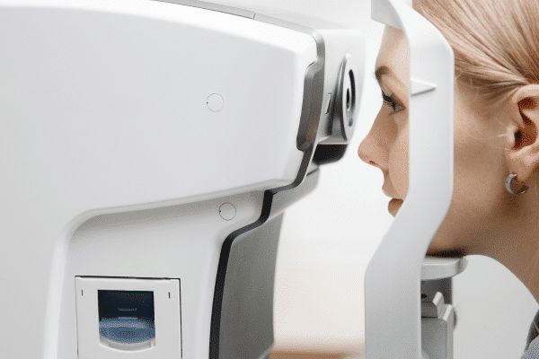
It is a completely non-invasive technique with which cross-sections of the retina, cornea and vitreous body can be imaged, and the optic nerve disc can be evaluated.
OCT vision testing emerged in ophthalmology in the 1990s. It was launched in the 1970s and revolutionized fundus imaging from the get-go. They are mainly performed in the diagnosis of macular disorders and in controlling the effectiveness of glaucomatous neuropathy treatment.
What does the OCT study consist of?
OCT examination (English: Optical Coherence Tomography) of the eye is a modern diagnostic method that allows detailed analysis of the eye’s structures in layers. It uses optical light technology to obtain precise images of the eye’s tissues.
During the OCT examination, the patient places his or her chin on a support, and a special OCT camera is placed in front of the eye. The patient is asked to direct his or her gaze to the fixation point, and the device then scans the eye, generating a detailed cross-sectional and vertical image.
OCT examination allows analysis of various structures of the eye, such as the retina, optic nerve, blood vessels and corneal layers. This allows the doctor to assess the thickness, density, possible lesions, damage or swelling of the tissues.
One of the main advantages of OCT examination is its non-invasiveness. It does not require dropping drops into the eye or special preparations. It is fast, precise and comfortable for the patient.
When should an OCT study be performed?
OCT examination of the eye is useful in many situations, both for diagnosis and for monitoring eye diseases. Among other things, this test is indicated for diagnosis and monitoring:
- Retinal diseases: OCT is particularly useful for retinal diseases such as macular degeneration (AMD), diabetic retinopathy, macular edema, retinoblastoma, and retinal dystrophies. It allows accurate imaging of retinal structures and evaluation of possible pathological changes.
- Vascular diseases: OCT can be used for vascular diseases of the eye, such as hypertensive retinopathy and central retinal artery occlusions. It allows assessment of possible blood vessel damage and intraretinal lesions.
- Optic Nerve Diseases: OCT is useful in diagnosing and monitoring diseases of the optic nerve, such as glaucoma and optic neuropathy. It allows to assess the structure of the optic nerve, the thickness of the nerve fibers and possible damage.
- Treatment undertaken: OCT examination allows monitoring the effectiveness of treatment for eye diseases. The doctor can assess whether the therapy is successful and adjust further treatment depending on the test results.
- New developments: Regular OCT examinations may be advisable to monitor eye health in people at risk of developing eye diseases, such as those with diabetes, the elderly or those with significant visual impairment.
If various diseases are suspected, an ophthalmologist will refer an eye doctor for this examination, while regular OCT examinations can contribute to early detection and effective treatment of various eye diseases, which is important for maintaining vision health.
Are you interested in eye examinations?
You are welcome to schedule a consultation.
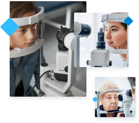
More than 120 years of experience passed down from generation to generation.
Expert content on our blog
July 26, 2022
Laser Vision Correction in Krakow – Vlasik Method
See what our proprietary vision correction method is all about.
Read
June 23, 2021
Refraction of the eye – what is refraction and what are the main refractive disorders of the eye?
What are refractive errors and how are they corrected?
Read
June 7, 2021
What eye defects qualify for a disability certificate?
When a visual defect makes normal life difficult or impossible, a patient can apply for a disability certificate. What eye defects qualify for a disability rating?
Read
