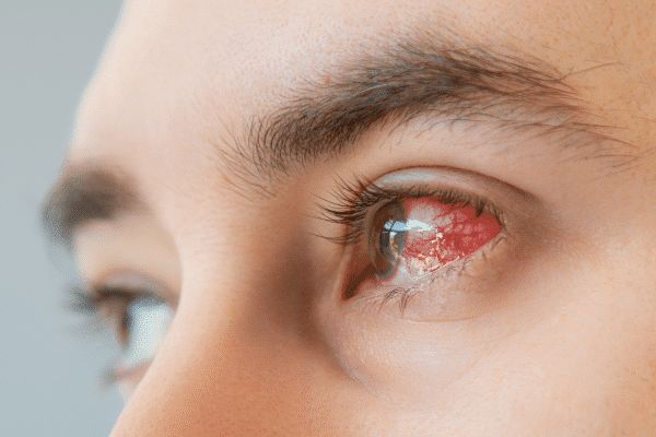
The cornea is the outer, convex layer of the eye, located at the front of the eye. It plays a very important role in the process of vision - it "transmits" light radiation to the eyeball and refracts it accordingly. For the mechanism to work properly, this part of the eye must be fully transparent. Corneal inflammation, on the other hand, can interfere with the transparency of the cornea, and thus the process of vision, so the disease must be treated as soon as possible.
Keratitis - types and kinds
Keratitis can have various causes. In this regard, infectious and non-infectious inflammation are distinguished.
In a disease caused by an infection, these will be:
- Bacterial keratitis - as the name suggests, it is caused by bacteria, for example, after damage to the corneal epithelium or in dry eye syndrome, where the organ of vision is not adequately protected due to too little tear film. Inflammation can be caused by bacteria such as Neisseria gonorrhoeae, Haemophilus influenzae, Staphylococus aureus, Pseudomonas aeruginosa,
- Fungal keratitis - is caused by fungi. It is often associated with contact with wood or the use of infected contact lens care solutions,
- Viral keratitis - is mainly encountered in immunocompromised individuals. In the case of keratoconjunctivitis, it can be caused by adenovirus types 8, 19 or 37. Another type of virus is Herpes zoster ophthalmicus , which causes ocular herpes zoster , or Keratitis herpetica , which causes herpetic keratitis,
- Protozoal keratitis - also known as creeping keratitis, is caused by Acanthamoeb, a single-celled creeper. It can be associated with inadequate hygiene when using contact lenses.
In non-infectious keratitis, the cause is sometimes an allergy, damage to the corneal innervation, an autoimmune disease or a systemic, or systemic disease.
The condition also distinguishes between its two forms - ulcerative keratitis when the epithelium is damaged and non-ulcerative, when there is no damage to the epithelium.
Keratitis - symptoms
Among the symptoms of keratitis, patients most often mention acute eye pain, photophobia, decreased visual acuity, redness of the eye, swelling of the eyelids and mucopurulent discharge in the conjunctival sac.
However, the symptoms vary depending on the type of inflammation:
- In a bacterial infection, this can include limited white corneal opacity, necrosis of the entire cornea and a very thick mucopurulent secretion.
- In fungal infection - the examination shows crater-like white-gray ulcers with a dry central base and fluffy white-colored frass.
- In viral keratitis - in the case of adenoviruses - patients complain of headaches, enlarged lymph nodes near the ears, severe redness of the conjunctiva, sometimes bloody petechiae and circular opacities on the cornea that interfere with vision. In ocular hemiplegia, the symptoms are pinpoint defects in the corneal epithelium, vesicles near the eye - on the eyelid, nose, forehead. In herpetic inflammation, characteristic tree-shaped epithelial defects appear.
Primary keratitis, on the other hand, is accompanied by severe pain when rubbing the eyelids and an annular haze of the cornea.
Keratitis - complications
Unfortunately, inflammation of this part of the eye can lead to complications. People who have undergone viral keratitis are particularly vulnerable to them.
The disease can cause corneal opacity, corneal ulceration, perforation, inflammation of the interior of the ocular tissue and even total inflammation of the cornea. Complications sometimes also involve disruption of the eye's architecture - once the inflammation has subsided, secondary glaucoma or cataracts appear. The risk of bacterial superinfection is also increasing. One of the most common complications that patients complain about is the deterioration of vision after keratitis. The reason for this condition is the scarring that resulted from the disease.
How are complications from keratitis treated?
Both glaucoma and secondary cataracts can be treated with a laser. In the case of glaucoma , it is a laser trabeculoplasty procedure aimed at lowering intraocular pressure.
In secondary cataracts , capsulotomy works well - the procedure, performed with a laser beam, takes just a few seconds. The procedure involves making a small hole in the center of the posterior lens capsule. Patients experience an immediate improvement in vision quality.
As for ulcers, the doctor uses antibiotics, steroid drops and therapeutic lenses. Corneal perforation, on the other hand, requires corneal transplantation as soon as possible.
Inflammation of the inside of the eye tissue involves the need for antibiotics and, if it progresses rapidly, a vitrectomy procedure that involves excising the vitreous body from inside the eye. This is usually the last chance to regain or maintain vision.
Treatment removal of scars after keratitis can be performed by laser. A very popular PTK procedure is the removal or reshaping of corneal fragments using photoablation. The device works only on the surface of the cornea, so there is no risk of damaging the inner layers. Laser corneal scar removal procedures are offered by our eye clinic in Krakow - Voigt Eye Clinic.

